The Spurlock Museum mummy gets a CT-scan
- Post Date: 5/20/2011
- Author: John Holton
- Reading Time: 4 minute read
For the second time since it arrived in Urbana in 1989, our ancient Egyptian mummy has made a trip to the hospital. The Spurlock Museum has joined forces with Carle Foundation Hospital, the Illinois State Archaeological Survey (ISAS), and the Smithsonian Institution to carry out a major new study of the Museum's oldest resident. On March 29, 2011, the mummy was taken to Carle Hospital, where it underwent a series of intensive CT-scans under the direction of Dr. Joseph Barkmeier.
This page includes a series of photographs that tell the story of preparing the mummy for its trip to the hospital, the taking of the CT-scans, and the return of the mummy to its case in the Museum, along with a peek at some of the new images.
-
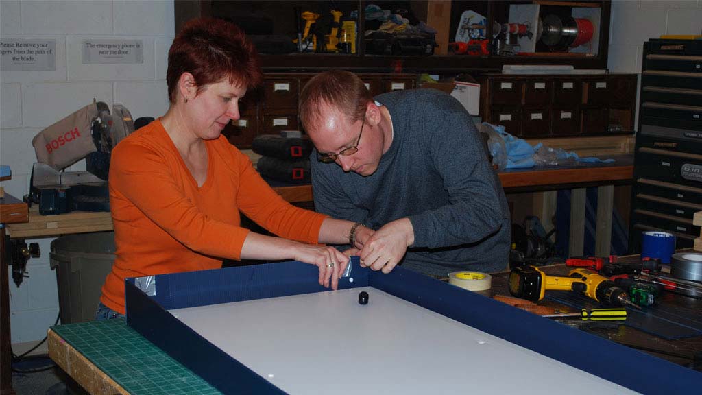 John Holton and Christa Deacy-Quinn install fasteners to the mummy crate's lid.
 -
 Holton and Deacy-Quinn place mummy into the crate.
 -
 Mummy inside its padded, custom crate.
 -
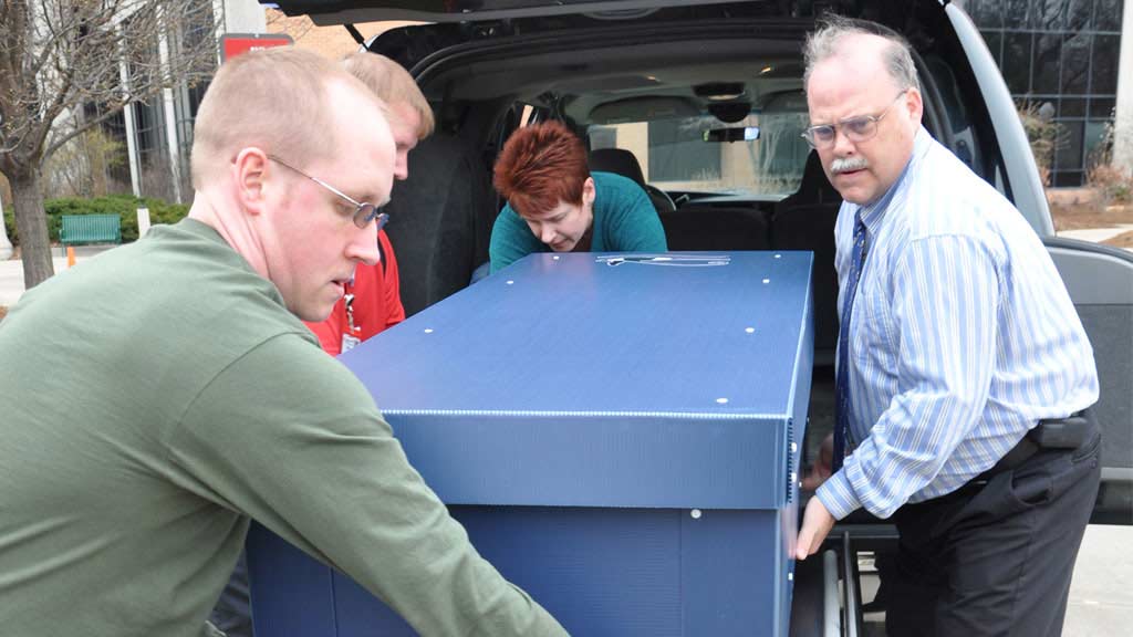 The mummy crate is transferred to a gurney at Carle Foundation Hospital. From the bottom left, clockwise; John Holton, assistant collections manager at Spurlock Museum; Christa Deacy-Quinn, collections manager; and David Hunt physical anthropology museum specialist and collections manager, Smithsonian National Museum of Natural History.
 -
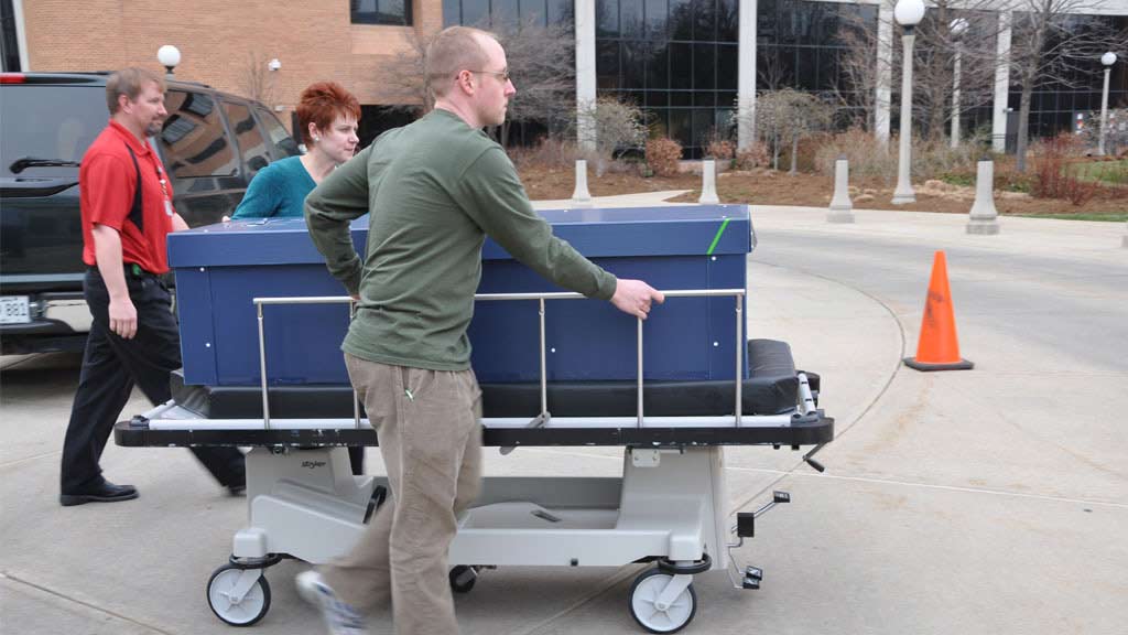 Mike Graybill (back) escorts Deacy-Quinn and Holton as they move the mummy on a hospital gurney.
 -
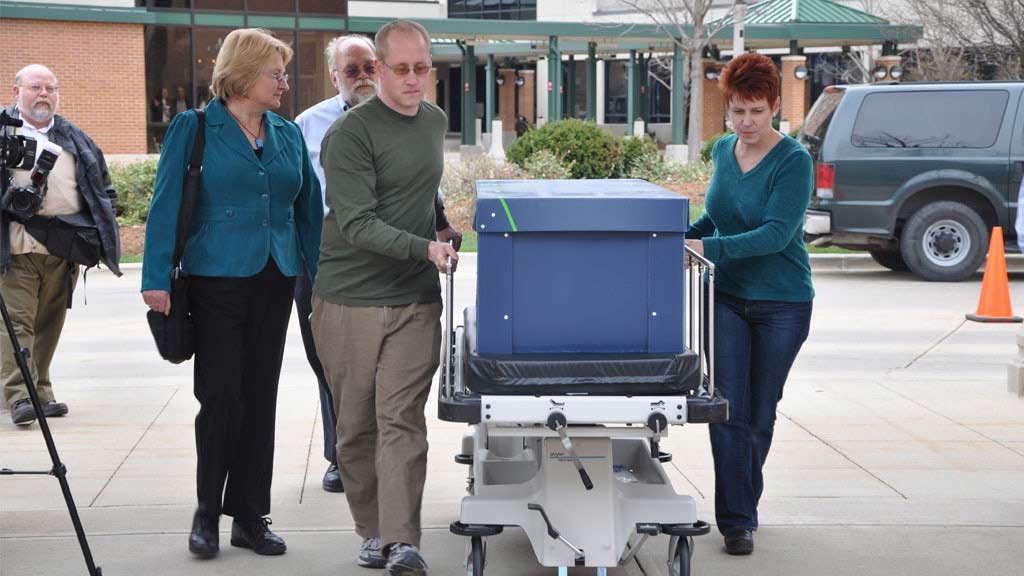 Sarah Wisseman (left) and Wayne Pitard (back) on-look as Holton and Deacy-Quinn move the mummy through the hospital entrance.
-
 Deacy-Quinn and Holton move the mummy to the CT scanning bed.
 -
 Graybill directs Deacy-Quinn and Holton as they make final adjustments before the scan begins.
 -
 Pitard (left), Joseph Barkmeier (middle), and Wisseman (right) discuss the mummy before it is scanned.
 -
 Laser sight lines are used to ensure the mummy is in the optimal scanning position.
 -
 The mummy rests on the table as the scan begins.
 -
 Full view of the mummy during the CT scan process.
 -
 The mummy's point of view as staff looks on.
 -
 The mummy waits as the scan completes.
A number of scholars, led by Dr. Sarah Wisseman of ISAS and Dr. David Hunt, a forensic anthropologist at the Smithsonian, will study the results over the next several months and will present their findings at a symposium at the Spurlock in November. Dr. Wisseman also organized the first study in the early 1990s, which showed that the mummy holds the remains of a young child between the ages of 7 and 9 who lived and died in the Fayyum Oasis in Egypt between 50 and 150 CE. But the original CT-scan did not enable scholars to discern whether the child was a boy or girl, and there was not enough evidence to determine a cause of death. CT-scan technology has improved dramatically over the past 20 years, and Wisseman and Hunt believe that we will gain considerably more information from the new data.
-
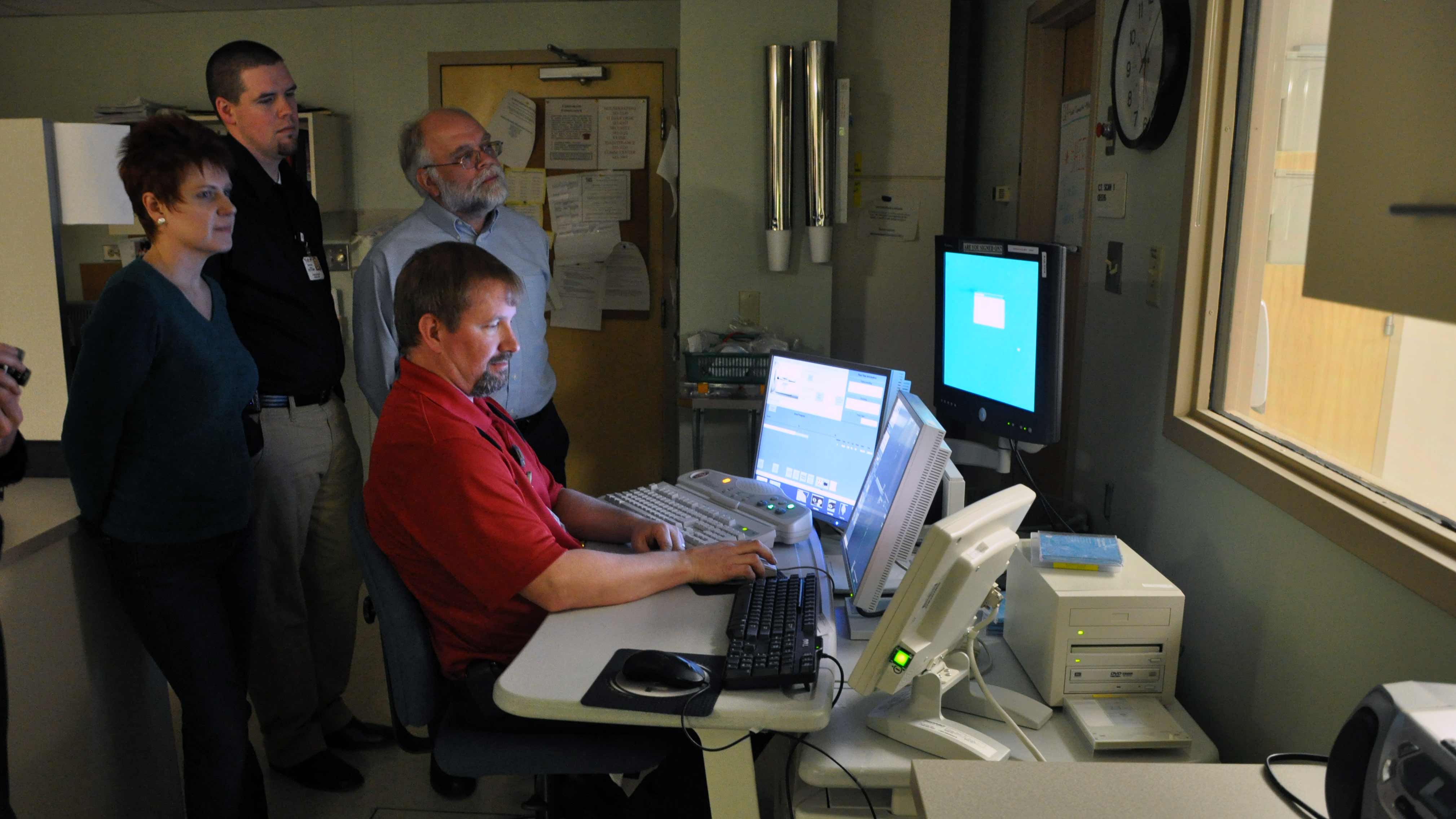
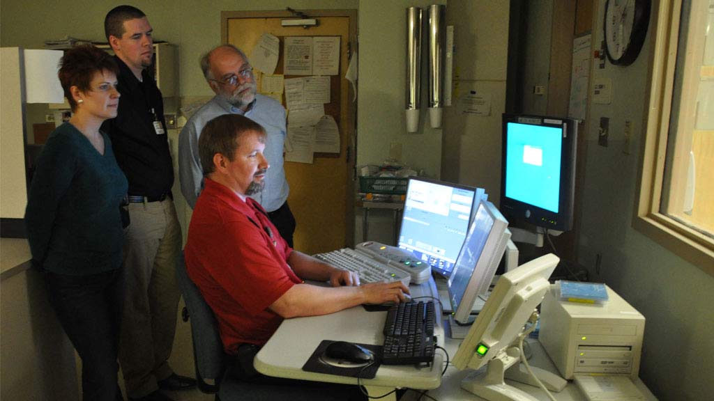 Mike Graybell (seated), Carle lead CT technologist, gathers data from the CT scanner. Monitoring the process of the CT scan: from left, Deacy-Quinn; Donovan Beswick, Carle radiology imaging manager; and Wayne Pitard, the director of the Spurlock Museum and a professor of religion.
 -
 CT imaging data as it appears on the screen during the scan.
 -
 Another view of the imaging data.
 -
 The mummy’s skeletal system can be easily seen in this image of the scan.
-
- Share:
- Subscribe to Newletter
- Giving